I have spots on my skin: where do I start to understand what they are?
The first step is not to use the word “spot” as though it were a diagnosis. A spot may be darker than the surrounding skin, lighter, isolated, diffuse or form a patch. Four pieces of information are especially useful for orientation: what the pattern looks like, where it is, when it appeared and how it behaves over time.
One isolated brown lesion on a highly sun-exposed area raises different questions from symmetrical pigmentation on both cheeks, a mark left after a pimple or an area of skin that has lost color. Before asking “how do I remove it?”, it is more useful to answer a more basic question: “what type of pigment change am I looking at?”.
What changes if the pigmentation is isolated, diffuse or appears in patches?
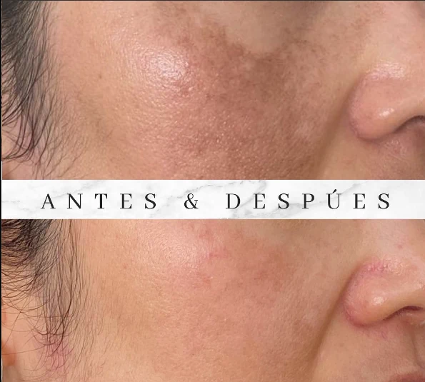
Distribution is one of the most useful clues. An isolated spot is a specific lesion that can be pointed to individually. A patch covers a broader area and may appear in a similar way on both sides of the face. Diffuse pigmentation makes an area look darker or uneven without a precise border.
This pattern does not allow self-diagnosis, but it helps organize the possibilities. solar lentigines are usually focal and individually countable. melasma tends to form broader areas, often symmetrically, and has a history of darkening and recurring. Post-inflammatory pigmentation usually follows the site of previous acne, dermatitis, a burn or another injury.
What does a light or white patch on the skin mean?
A light patch changes the reasoning completely because we are no longer talking about excess pigment but about hypopigmentation or loss of color. It can occur after inflammation, with conditions such as vitiligo, with some superficial infections or for other reasons that need to be distinguished.
The important point is not to try to “even out” a white area as though it were a brown spot. If the loss of color is new, spreading, appearing in other areas or has no clear cause, the first step is to identify why that skin has less pigment.
How can a sun spot be distinguished from melasma?
An isolated sun-related spot is usually described as a small brown macule or patch in an area with accumulated exposure: the face, backs of the hands, arms or décolletage. When it corresponds to a solar lentigo, it remains an individual lesion.
melasma, by contrast, usually covers broader areas of the face, may involve the cheeks, forehead, nose or upper lip, and often appears in a similar pattern on both sides. It also tends to have periods when it darkens and others when it fades. The fact that both types of pigmentation are brown does not mean they have the same origin or should be managed in the same way.
Why can a dark mark remain after a pimple, irritation or injury?
Inflammation can stimulate pigment production. A pimple, dermatitis, burn, irritating hair removal, wound or procedure that left the skin inflamed can therefore leave a residual dark mark after the active episode has ended.
In that situation, the history matters greatly: if the mark appeared exactly where inflammation occurred, that relationship is more informative than color alone. It also explains why continuing to irritate the area can prolong or intensify the problem.
Why do spots appear with sun exposure, hormones or inflammation?
There is no single cause of skin pigmentation. Cumulative radiation can promote pigmented lesions in exposed areas; hormonal changes can act as triggers in people predisposed to melasma; and inflammation can leave pigment even when the sun was not the original cause.
Several factors can also coexist. A person may have photoaging, a few lentigines and post-inflammatory marks at the same time. The purpose of triage is not to force one universal explanation, but to separate the different components that are present.
Why do facial spots become more common with age?
“Age spots” is a useful everyday expression for what many people see, but it is not a diagnosis. Over the years, sun exposure accumulates and different signs of photoaging become visible, including solar lentigines and a more uneven skin tone.
Aging also does not explain every spot that appears after 40 or 50. Melasma, post-inflammatory pigmentation, keratoses and other lesions may coexist at that age. “It appeared because of age” is therefore a starting point, not a conclusion.
Does location matter: cheeks, forehead, upper lip, hands or décolletage?
Yes. Location changes the probabilities. The face, hands and décolletage accumulate sun exposure and commonly show photoaging-related changes. Melasma favors the face and may involve the cheeks, forehead, nose or upper lip. A post-inflammatory mark appears where the earlier inflammation occurred.
Location alone is never enough: a shadow above the upper lip is not automatically melasma, and a spot on the hand is not automatically a lentigo. Pattern, texture, history and evolution all need to fit together.
What does it mean if a spot darkens in summer, changes or comes back?
The relationship with seasons provides useful information. Melasma often has periods of greater and lesser intensity and can recur after improving. Lesions related to sun damage can become more visible with renewed exposure, and new spots can also appear over time.
“It came back” does not always mean the exact same lesion returned. Sometimes the same pigmentation pattern reappears; other times new nearby lesions develop. Distinguishing those situations helps clarify whether this is a recurrent condition or an accumulation of different changes.
What does uneven tone mean when there is no clearly defined spot?
Not every pigment alteration forms a round, easy-to-point-to lesion. Some people seek consultation because the face looks dull, with darker areas, scattered spots and generally uneven tone. In that situation, photoaging, residual pigmentation and other causes of hyperpigmentation may coexist.
When several things are present at once, giving them one name can be misleading. A person may have isolated lentigines as well as a different diffuse pattern. Assessment helps separate these components before deciding what should be addressed.
If you also have wrinkles, where should you start?
The first step is to identify what type of pigmentation is present, especially if any lesion is new, different or has changed. That does not mean pigmentation always has to be treated first: once anything requiring diagnosis has been ruled out, aesthetic priorities depend on what has the greatest impact on that face—uneven tone, lines, texture or a combination.
If your question is specifically what makes a face look older when pigmentation and wrinkles coexist, we address that angle in Do dark spots age the face more than wrinkles?.
Is a new brown spot always just an aesthetic concern?
No. Many pigment changes are benign, but a pigmented lesion should not be assumed to be cosmetic simply because it is brown. If a spot is new and different from the others, changes in size, shape or color, contains several colors, bleeds, ulcerates or evolves in a way that draws attention, lightening it is not the priority: evaluating it is.
When an individual lesion does not fit a common benign pattern, dermatologic assessment—and dermoscopy when appropriate—may be needed before any aesthetic intervention.
Why is it a bad idea to try to lighten a spot before knowing what it is?
Because “dark” describes a color, not a cause. An overly irritating strategy can add inflammation and post-inflammatory pigmentation. And if the lesion has not yet been properly identified, trying to erase it can delay an assessment that may be necessary.
The safer sequence is simple: classify first, decide second. Before choosing a product, acid or technology, it is useful to understand whether the pattern is focal, diffuse, recurrent, post-inflammatory or represents a lesion that needs a different type of evaluation.
Can you tell what type of pigmentation it is from a photograph alone?
A photograph can provide clues, but it has limits. Lighting changes color, filters alter contrast, and an image does not tell you whether the spot appeared after pregnancy, followed dermatitis, darkens every summer or has been changing for months.
Distinguishing pigment patterns usually requires combining appearance + distribution + history + evolution. And when an individual lesion raises questions, an in-person examination provides information that a photograph cannot.
What is useful to observe before a consultation for pigmentation?
You do not need to arrive with a diagnosis. It does help to reconstruct the history:
- when you first noticed the pigmentation;
- whether it appeared suddenly or gradually;
- whether there was pregnancy, hormonal change, medication, acne, dermatitis, irritation or a previous injury;
- whether it worsens at certain times of year;
- whether it changed in size, shape, texture, border or color;
- which products or procedures you used and how the skin reacted;
- whether there are similar spots on the face, hands, arms or décolletage.
These details turn “I have spots” into a much more useful description of the problem.
When is it worth seeking an assessment, and what should a medical evaluation resolve?
An assessment is useful when you do not know what type of pigmentation you have, when it is increasing, recurring, when several types coexist, or when an individual lesion has changed and raises concern.
A good first assessment should answer: what pattern is this?, which factors are maintaining it?, is more than one cause present?, does it look more like lentigines, melasma or another process?, is there any lesion that needs dermatologic evaluation before aesthetic treatment?
That initial diagnosis is the starting point for the cluster: if focal sun-related spots predominate, you can read more about solar lentigines; if recurrent facial patches predominate, continue to melasma.
The fact that a technology exists does not mean it is indicated for your pigmentation
Q-Switch, intense pulsed light, peels and other procedures may have specific indications, but not every type of pigmentation is a candidate for the same options. The first step is to identify the pattern, assess skin phototype, understand how the pigmentation behaves and rule out lesions that need another type of evaluation.
Once the pigmentation has been correctly classified, some technologies may enter the discussion depending on the case. The cards below explain those options; they are not automatic recommendations.