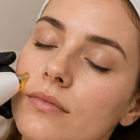
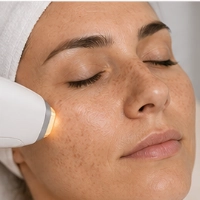
I have small brown spots: could they be solar lentigines?
Solar lentigines usually appear as flat, brown, relatively well-defined spots on skin that has received sun exposure for years. They are commonly seen on the face, backs of the hands, forearms, shoulders and décolletage.
That appearance is suggestive, but it does not confirm by itself that a lesion is a lentigo. Other benign spots—and some lesions that require evaluation—can look similar to the naked eye. The first useful question is “does this lesion really have the characteristics of a solar lentigo?”.
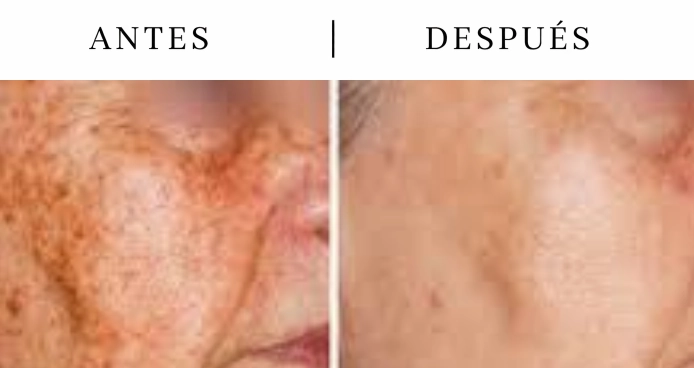
What exactly is a solar lentigo?
It is an acquired pigmented lesion associated with accumulated sun exposure. Unlike diffuse pigmentation, a lentigo can be identified as an individual lesion: you can point to where it begins and ends, even if its border is not a perfect circle.
It is usually asymptomatic, and there may be one lesion or many. When numerous lentigines are present, the skin can look generally more uneven even though each spot remains focal.
Why do they appear now if the sun damage happened years ago?
Visible changes do not necessarily appear at the same time as the exposure that contributed to them. Radiation accumulated over years creates changes that can become evident gradually. Someone may therefore begin to notice lentigines in adulthood even if the period of greatest sun exposure occurred much earlier.
The skin does not “reset” its history each season either: the current pattern reflects part of what happened over many years as well as what continues to happen.
Why do solar lentigines become more common with age?
Age itself does not directly create them. What increases over the years is the cumulative time of exposure and the opportunity for photoaging to become visible. That is where the popular term “age spots” comes from, even though they do not appear simply because someone has had another birthday.
Two people of the same age can have very different numbers of lentigines because of their exposure history, skin phototype, individual predisposition and sun-protection habits.
Where do solar lentigines appear most often?
They predominate on chronically exposed areas: the face, hands, forearms, shoulders and décolletage. Distribution provides a clue because a collection of focal spots on exposed skin fits solar lentigines better than broad, symmetrical pigmentation limited to the central face.
Even so, location alone is not enough for diagnosis. The appearance of each lesion and how it changes over time still matter.
Spots on the hands: why are solar lentigines so common there?
The backs of the hands receive radiation almost every day—while walking, driving, doing outdoor activities or simply being near windows. Repeated exposure helps explain why solar lentigines are so common in this area.
However, not every brown spot on the hand is a lentigo. Adults may also develop seborrheic keratoses, pigmented actinic keratoses, moles and other lesions. If one spot looks different from the others or is changing, it should be identified first.
Is a lentigo the same as a freckle?
Not exactly. Freckles usually appear at younger ages and tend to become more visible with sun exposure and fade when exposure decreases. Solar lentigines are acquired lesions that generally appear later and persist.
To the naked eye they may share a similar color and size, so in a person with many pigmented lesions it can be difficult to label every spot without assessment.
How can solar lentigines be distinguished from melasma?
The most useful difference is the pattern. Lentigines are usually focal spots that can be counted individually. melasma usually forms broader areas of facial pigmentation and is often distributed in a similar way on both sides of the face.
The history also differs: melasma commonly darkens, lightens and recurs, and may be associated with hormonal factors. Solar lentigines fit better with accumulated exposure and photoaging. If you are still not sure which pattern you are describing, return to the skin pigmentationoverview.
How can a lentigo be distinguished from a seborrheic keratosis, a mole or another pigmented lesion?
A flat seborrheic keratosis can look very similar to a solar lentigo. Moles and other pigmented lesions may also share a similar color or size. Texture, elevation, regularity, evolution and, when necessary, dermoscopy help distinguish them.
This matters because not every brown spot should be treated as cosmetic pigmentation. A lesion that looks different from the others, changes in a striking way or has an uncertain diagnosis needs assessment before anyone tries to alter its appearance.
Can a lentigo grow, darken or appear together with other spots?
Lentigines can become more noticeable over time, and new lesions can appear on exposed skin. That does not mean every change should automatically be attributed to a “normal” lentigo. A lesion that changes in an unusual or striking way deserves review.
It is also common for different types of pigmented lesions to coexist in the same area. Assessment is not about giving the same label to everything that is brown.
Do solar lentigines disappear on their own or change between winter and summer?
An established solar lentigo usually persists. Its visibility can vary depending on skin contrast and exposure, but it does not typically behave like a freckle that fades clearly during months with less radiation.
And even when one particular lesion remains stable, new ones can appear. The feeling that “I have more every year” often reflects this gradual accumulation.
Does having many lentigines mean the skin has had too much sun?
Solar lentigines are a visible sign of accumulated exposure and photoaging. They cannot calculate how much radiation a person has received and do not by themselves predict what will happen to the skin, but they do tell part of that history.
That is why, when many lesions are present on exposed areas, it is useful to look beyond the single spot that is bothersome and assess the overall condition of the skin and whether other sun-related changes are present.
Can solar lentigines be removed?
Many correctly identified solar lentigines can lighten substantially and may even become no longer visible, but the result depends on the lesion, its color, skin phototype, the number of spots and the individual skin response. New lentigines may also develop over the years even after previous ones have improved.
The most important step comes first: confirming that the lesion really is a benign lentigo. Once that diagnosis is established, different ways of treating solar pigmentation can be considered. In selected patients, one of the options we use for sun spots is intense pulsed light (IPL).
The link explains the technology; it does not mean every person with lentigines should receive IPL. Indication depends on the pattern of spots and assessment of the skin.
What can be expected from solar lentigines as the years go by?
The general tendency is for new lesions to appear as exposure and skin aging accumulate. Some remain quite stable; others become more visible. This explains why someone may feel that pigmentation is “progressing” even when no individual spot has changed in a concerning way.
Periodic assessment of exposed skin helps separate expected changes from those that deserve a closer look.
What can be learned from a photograph, and what is useful to observe before a consultation?
An image can show whether a lesion appears focal and pigmented, but it does not reproduce texture well and cannot tell how the lesion evolved. Color also changes depending on camera and lighting.
Before a consultation, it helps to remember whether the spot has been present for years or appeared recently, whether it changed in size or color, whether there are similar spots nearby, how much sun that area received, and whether it has ever bled, crusted or caused symptoms. Older photographs can be useful for comparison.
When does a medical assessment for lentigines make sense?
It is useful when you are not sure that the spots are truly lentigines, when there are many lesions that look different from one another, when one has changed, or when you want to separate pigmentation caused by photoaging from another condition.
The first step is not deciding how to erase the spot. It is to confirm that the lesion is compatible with a solar lentigo and distinguish it from other pigment changes. If the assessment confirms lentigines and the goal is to reduce them, then it makes sense to discuss options such as intense pulsed light.
The fact that a technology exists does not mean it is indicated for your lentigines
Intense pulsed light can be part of the management of selected solar lentigines, but it should not be indicated simply because a spot looks like an “age spot.” First, the lesion should be confirmed as a benign pattern compatible with a lentigo, skin phototype and number of lesions should be assessed, and spots requiring a different type of evaluation should be excluded.
If the diagnosis confirms solar lentigines and the goal is to reduce their contrast, the next page explains the technology we use as the main pathway from this concern.